Neck Anatomy Diagram : Neck Anatomy And Physiology Elliot S Site
Neck Anatomy Diagram : Neck Anatomy And Physiology Elliot S Site. The thyroid gland is located in the neck below the thyroid cartilage, or adam's apple. Contains cervical vertebrae and postural muscles. Muscles of the neck (musculi cervicales) the muscles of the neck are muscles that cover the area of the neck hese muscles are mainly responsible for the movement of the head in all directions they consist of 3 main groups of muscles: Know your spine, neck pain. The purpose of the spine is to support the body so that we can stand upright.
ads/bitcoin1.txt
Learn the anatomy of the compartments of the neck with this quiz, video, articles a. Anterior, lateral and posterior groups, based on their position in the neck.the musculature of the neck is further divided into more specific groups based. The head and neck is covered in skin and its appendages, termed the integumentary system.these include hair, sweat glands, sebaceous glands, and sensory nerves.the skin is made up of three microscopic layers: In addition, in this region we also find the major cranial and spinal nerves that connect the central nervous system to the organs, skin, and muscles of the head and neck. 8 ways to help protect your cervical spine.
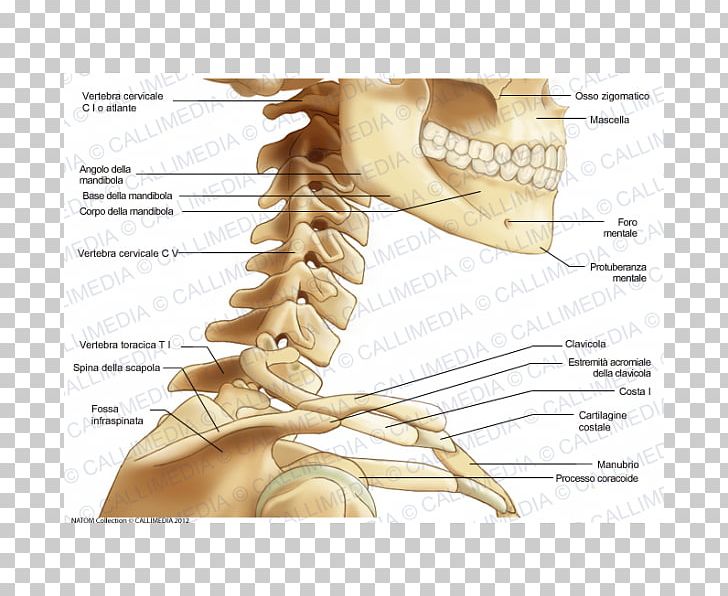
Bone Human Skeleton Neck Anatomy Png Clipart Anatomy Arm Bone Claw Ear Free Png Download from cdn.imgbin.com It is important to note that all triangles mentioned here are paired; Clinically, surface anatomy is used to split the neck into anterior and posterior triangles which provide clues as to the location of specific structures. There are 33 bones in the spine. The neck muscles, including the sternocleidomastoid and the trapezius, are responsible for the gross motor movement in the muscular system of the head and neck. Very soon we'll move on to muscles! Learn more about the anatomy of the neck in this section. Almost every bone in your body is made of the same materials: The anatomy of the head and neck of the human body, including the bones, muscles, blood vessels, nerves, glands, nose, mouth, and throat.
Anterior, lateral and posterior groups, based on their position in the neck.the musculature of the neck is further divided into more specific groups based.
ads/bitcoin2.txt
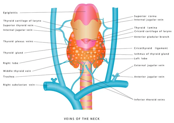
The external jugular vein (v. In this video, we will cover the anterior muscles of the neck, and talk about their origins, insertions, innervation and functions. Anatomy of the neck the neck contains a number of overlapping muscles blood vessels nerves and interactive anatomical atlas of the head, brain, and neck based on anatomical diagrams and ct and. Learn more about the anatomy of the neck in this section. There are two long bones in your arm which are connected through. Contains glands ( thyroid , parathyroid, and thymus ), the larynx , pharynx and trachea. Neck neck muscle anatomy muscle diagram inspirational medical. Shoulder girdle , radiographs :. Neck pain is a common symptom, although not all patients experience pain. The parts of the body where people and their doctors can see or feel swollen lymph nodes include the neck, armpit, and groin areas. The thyroid gland is located in the neck below the thyroid cartilage, or adam's apple. The purpose of the spine is to support the body so that we can stand upright. The cervical spine is delicate—housing the spinal cord that sends messages from the brain to control all aspects of the body—while also remarkably strong and flexible, allowing the neck to move in all directions.
Causes of neck pain and how to manage the pain in basic terms, the neck (cervical spine) joins the shoulders and chest to the head. Diagram of bones in neck and shoulder / neck anatomy pictures bones muscles nerves : The anterior triangle is a region located at the front of the neck. The cervical spine is delicate—housing the spinal cord that sends messages from the brain to control all aspects of the body—while also remarkably strong and flexible, allowing the neck to move in all directions. Veins and arteries of the neck 9 photos of the veins and arteries of the neck activate javascript arteries in the neck diagram, common carotid artery branches, external carotid artery function, how many carotid arteries, left common carotid artery function, the left common carotid artery supplies blood to the.
Head And Neck Anatomy Png Images Pngwing from w7.pngwing.com Related posts of diagram of the neck anatomy veins and arteries of the neck. Learn everything about the neck anatomy with this topic page. Jugularis externa) receives the greater part of the blood from the exterior of the cranium and the deep parts of the face, being formed by the junction of the posterior division of the posterior facial with the posterior auricular vein.it commences in the substance of the parotid gland, on a level with the angle of the mandible, and runs perpendicularly down the. Causes of neck pain and how to manage the pain in basic terms, the neck (cervical spine) joins the shoulders and chest to the head. By sally ann quirke, chartered physiotherapist | filed under: They move the head in every direction, pulling the skull and jaw towards the shoulders, spine, and scapula. Learn the anatomy of the compartments of the neck with this quiz, video, articles a. Shoulder joint of human body anatomy infographic diagram with all parts including bones ligaments muscles bursa cavity capsule cartilage membrane for medical science education and health care.
Surface anatomy and surface markings bibliographic record list of illustrations the external jugular vein varies in size, bearing an inverse proportion.
ads/bitcoin2.txt
Neck pain is a common symptom, although not all patients experience pain. The neck contains seven of. It is extremely important because every cell in the body depends on the hormones the thyroid produces to. The neck is the start of the spinal column and spinal cord. The muscles of the neck run from the base of the skull to the upper back and work together to bend the head and. Muscles of the neck (musculi cervicales) the muscles of the neck are muscles that cover the area of the neck hese muscles are mainly responsible for the movement of the head in all directions they consist of 3 main groups of muscles: 8 ways to help protect your cervical spine. The content of the neck is grouped into 4 neck spaces, called the compartments. The content of the neck is grouped into neck spaces, called compartments. The largest vein in the neck is usually the internal jugular vein, which drains blood from the brain, neck muscles, face and organs of the neck. Know your spine, neck pain. The internal jugular vein commences at the jugular foramen, and is the direct continuation of the sigmoid sinus, which is a large vein draining blood from the vein. The nerves of the head and neck include the most vital and important organs of the nervous system — the brain and spinal cord — as well as the organs of the special senses.
In addition, in this region we also find the major cranial and spinal nerves that connect the central nervous system to the organs, skin, and muscles of the head and neck. In this video, we will cover the anterior muscles of the neck, and talk about their origins, insertions, innervation and functions. In radiology, the 'head and neck' refers to all the anatomical structures in this region excluding the central nervous system, that is, the brain and spinal co. They are located on both the left and the right sides of the neck. There are 33 bones in the spine.
Neck Anatomy Images Search Images On Everypixel from st2.depositphotos.com Almost every bone in your body is made of the same materials: The internal jugular vein commences at the jugular foramen, and is the direct continuation of the sigmoid sinus, which is a large vein draining blood from the vein. Anatomy of back of human neck, anatomy of the back and neck, anatomy of the back of the neck, anatomy of the back of the neck muscles, anatomy of the back of your. The thyroid gland is located in the neck below the thyroid cartilage, or adam's apple. The cervical spine is delicate—housing the spinal cord that sends messages from the brain to control all aspects of the body—while also remarkably strong and flexible, allowing the neck to move in all directions. There are two long bones in your arm which are connected through. The head and neck is covered in skin and its appendages, termed the integumentary system.these include hair, sweat glands, sebaceous glands, and sensory nerves.the skin is made up of three microscopic layers: The largest vein in the neck is usually the internal jugular vein, which drains blood from the brain, neck muscles, face and organs of the neck.
The head and neck is covered in skin and its appendages, termed the integumentary system.these include hair, sweat glands, sebaceous glands, and sensory nerves.the skin is made up of three microscopic layers:
ads/bitcoin2.txt
The neck is the start of the spinal column and spinal cord. The cervical spine is delicate—housing the spinal cord that sends messages from the brain to control all aspects of the body—while also remarkably strong and flexible, allowing the neck to move in all directions. Anterior, lateral and posterior groups, based on their position in the neck.the musculature of the neck is further divided into more specific groups based. Shoulder girdle , radiographs :. Understanding the anatomy of your cervical spine and the vital nerves it contains should motivate you to adopt behaviors that help prevent neck injury and slow development of degenerative disorders (eg, cervical disc herniation). Contains cervical vertebrae and postural muscles. The neck muscles, including the sternocleidomastoid and the trapezius, are responsible for the gross motor movement in the muscular system of the head and neck. The content of the neck is grouped into neck spaces, called compartments. Neck pain is a common symptom, although not all patients experience pain. The content of the neck is grouped into 4 neck spaces, called the compartments. Atlas of the anatomy of the head and neck on a ct in axial, coronal, and sagittal sections, and 3d images Learn more about the anatomy of the neck in this section. The muscles of the neck run from the base of the skull to the upper back and work together to bend the head and.
ads/bitcoin3.txt
ads/bitcoin4.txt
ads/bitcoin5.txt
0 Response to "Neck Anatomy Diagram : Neck Anatomy And Physiology Elliot S Site"


0 Response to "Neck Anatomy Diagram : Neck Anatomy And Physiology Elliot S Site"
Post a Comment